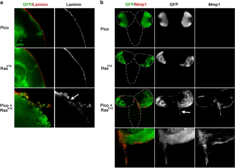
Figure 2.
Brains coexpressing RasV12 and pico display extracellular matrix degradation and ectopic expression of Mmp1. (a) Optic lobes from larvae overexpressing pico, RasV12 or RasV12, pico under the control of eyFLP, Act>GAL4, stained with anti-Laminin antibody, which labels the surface of the optic lobes. Laminin staining was found to be severely interrupted in brains coexpressing RasV12 and pico but not from brains expressing pico or RasV12 alone. (b) Distribution of the metalloproteinase Mmp1. Little or no Mmp1 staining was observed in animals expressing RasV12 or pico alone. In contrast, animals co-overexpressing RasV12 and pico had elevated Mmp1 around the edges of the optic lobes and at sites of invasion into the VNC (arrow).

