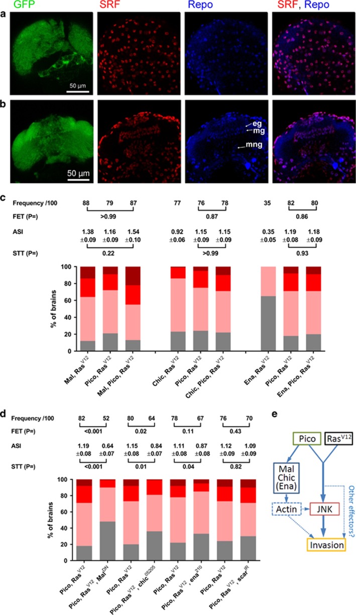
Figure 7.
Mal and Chic are rate limiting for tumour dissemination. (a, b) Distribution of SRF in the third instar larval optic lobes. SRF antibody staining (red) overlaps that of Repo (blue) both at the surface (a) of the optic lobes and in cross-section (b). In cross-section, staining in epithelial (eg), marginal (mg) and medulla neurophil (mng) glia is evident.(c) Cooperation between RasV12 and cytoskeletal regulators results in cancer cell invasion into the VNC. Stacked bar charts summarizing extent of invasion in the different genotypes (n=100 brains of each type) according to the scale introduced in Figure 1, with 3 being the most severe and 0 corresponding to no invasion. Shown above each group of charts is a summary of statistical tests for key pairwise combinations, as indicated, in each experiment. Frequency represents number of brains showing invasion/total number (100 in each case); FET, Fisher’s exact test; ASI, average stage score of invasion; STT, Student’s t-test. (d) malDN and chic loss of function partially suppress the effect of RasV12 pico co-overexpression. ena and scar loss of function have reduced or no effect, respectively. Results are grouped by sibling pairs (overexpressed pico RasV12±genetic modifier) and are displayed as in (c). (e) Model of cooperation between Pico and RasV12 co-overexpression. Pico and RasV12 cooperate to activate JNK, which is necessary for invasion of glia from the optic lobes into the VNC. Mal, Chic, and to a lesser extent Ena, are rate limiting for invasion. These regulators may contribute directly or indirectly to invasion via changes in the actin cytoskeleton (see Discussion).

