Figure 4.
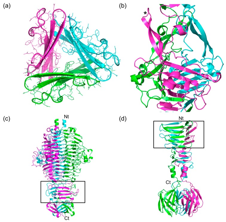
Triangular domains in bacteriophage fibers and tailspikes. (a) Cross-section of the P4 domain, viewed from the N-terminal end. (b) Side view of the P5 domain. One of the β-hairpins is indicated with an asterisk. The β-strands of the central β-sheet are numbered. Note that the fifth strand is irregular. (c) Side view of the bacteriophage P22 tailspike, which is a trimer of gp9. The triangular domain in this and the next panel are boxed and the termini are labelled. (d) Side view of the C-terminal part of the bacteriophage T7 tail fiber (gp17). In panels (c) and (d), N- and C-termini are labelled with Nt and Ct, respectively.

