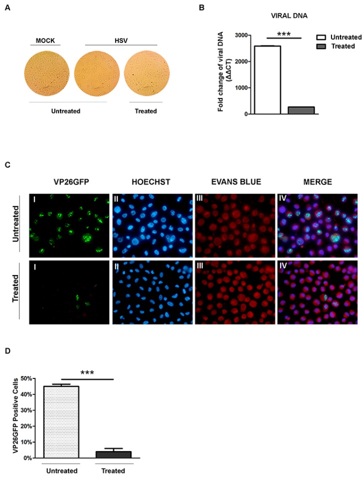
Figure 5.
Effect of almond extract treatment on the HSV-1 binding. Vero cells infected and mock-infected with HSV-1 at low temperature and processed as described in the Material and Methods. (A) Normal phase contrast inverted micrographs of processed samples are shown; (B) Relative quantization of viral DNA of the processed samples are shown. Values represent ±SD of the average of three samples normalized against the GAPDH copies number and were analyzed by the comparative Ct method (ΔΔCt); (C) Fluorescent images of Vero cells infected with VP26GFP HSV-1 virus and following virus inactivation, untreated and treated with NS extract. The green dots (I) represent VP16GFP viral antigen localization. The cells were stained with Hoechst (II) for the nuclear compartment and Evans blue dye (III) as a counter-stain. The VP26GFP, Hoechst and Evans blue images are merged in the last columns (IV); (D) Schematic representation of the VP26GFP-positive cells. Asterisks (***) indicate significant changes of p-values less than 0.001.

