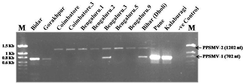
Figure 3.
Multiplex-RT-PCR for detection and differentiation of PPSMV-1 and PPSMV-2 isolates from SMD-affected pigeonpea samples. The lanes marked as “M” on the left and right extremes of the gel are the 100 bp DNA size marker (G Biosciences, St. Louis, MO, USA) for which the lengths of each bands are indicated. The two arrows point to the PPSMV-1 and PPSMV-2 specific amplicons and their sizes are also given.

