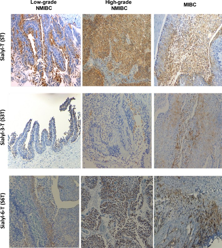
Figure 2.

Immunohistochemistry for sialylated T antigens (ST: corresponding to mono‐ and disialylated T glycoforms; S3T and S6T) for low‐ and high‐grade superficial papillary muscle‐invasive bladder tumours. The figure highlights the increase in T sialylation with the severity of the lesions. As the S6T antigen was determined based on comparisons with STn expression after β‐(1,3)‐galactosidase digestion, only STn‐negative tumour lesions are being presented in this figure. Moreover, because the S3T antigen expression was determined based on comparisons with T antigen expression after α‐(2,3)‐neuraminidase treatment, only T‐negative tissues are being presented.
