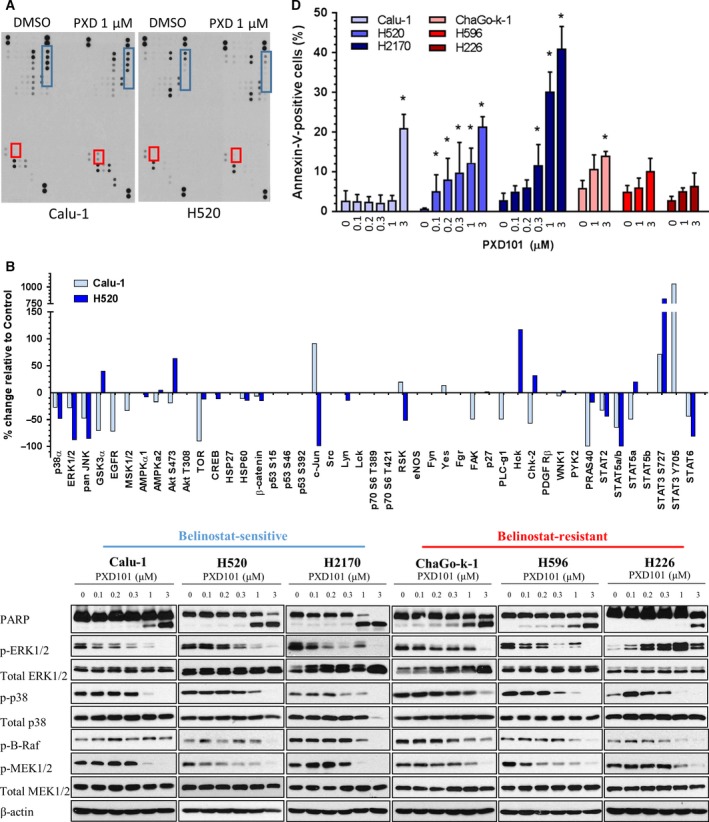
Figure 2.

Belinostat suppresses MAPK signaling and triggers apoptosis in lung squamous cell carcinoma (SCC). (A) Calu‐1 and H520 cells were treated with vehicle or 1 μm belinostat (PXD101) for 48 h with the protein lysates harvested for phosphokinase profiling. Dots representing consistently elevated targets are highlighted in red (STAT3), and suppressed kinases are highlighted in blue (p38, ERK1/2, JNK). (B) Densitometry analysis on the phosphokinase profiling was performed as described in Materials and methods. The average values of the significantly regulated duplicate were shown. (C) Cell lysates were harvested from lung SCC cells after treatment with increasing doses of belinostat (0.1, 0.2, 0.3, 1, 3 μm). Immunoblotting was performed to evaluate the changes in phosphorylated protein levels of the targets identified in 1D (ERK1/2, p38, B‐Raf, MEK1/2) as well as PARP. β‐Actin shown as loading control. (D) Lung SCC cells were treated with vehicle or belinostat (0.1, 0.2, 0.3, 1, 3 μm for Calu‐1, H520, and H2170; 1, 3 μm for ChaGo‐k‐1, H596, and H226) and stained with Annexin‐V/propidium iodide (PI). The percentage of Annexin‐V‐positive cells was shown as mean ± SD. *P < 0.05.
