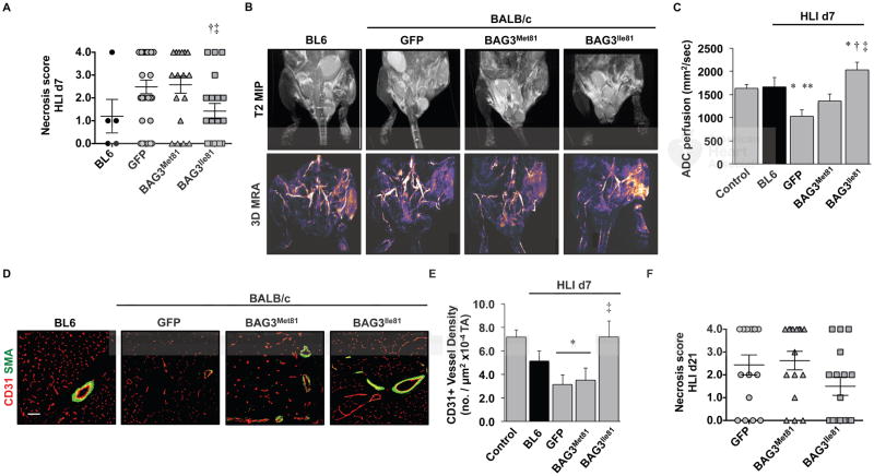
Figure 3. BAG3Ile81 expression regulates ischemic limb tissue necrosis and perfusion.
BL6 (N=5) and BALB/c mice were injected IM with AAVs (N=29 GFP; N=19 BAG3Met81; N=19 BAG3Ile81) and 7 days later subjected to HLI. A. Semi-quantitative scoring of limb muscle necrosis. †P<0.05 vs. GFP; ‡ P<0.05 vs. BAG3Met81. B. Representative MR T2-weighted and MR angiography images and (C) quantification of MR ADC perfusion at HLI d7 (N=4 BL6; N=8 GFP; N=7 BAG3Met81; N=6 BAG3Ile81). *P<0.05 vs. Control; **P<0.05 vs. BL6; †P<0.05 vs. GFP; ‡ P<0.05 vs. BAG3Met81. D. Representative images of CD31 and SMA staining (scale bar=100μm) (D) for quantification of CD31+ vessel density (E) at HLI d7 (N≥4 mice/group). *P<0.05 vs. Control; ‡ P<0.05 vs. GFP or BAG3Met81. F. Virus-injected BALB/c mice (N=16 mice/virus) were subjected to HLI for 21d, and limb necrosis score data plotted per mouse, with means ± SEM. All other data are means ± SEM.

