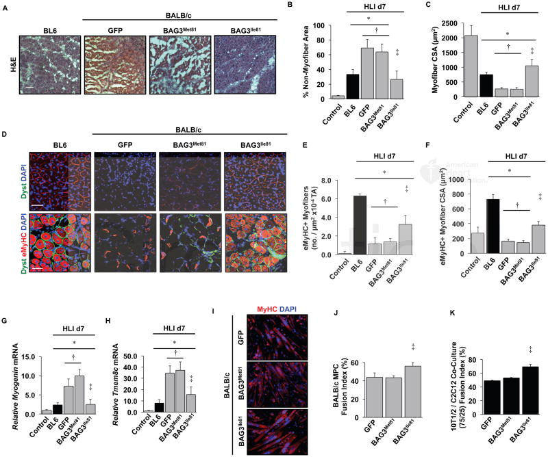
Figure 4. BAG3Ile81 rescues ischemic BALB/c muscle morphology and regeneration.
BL6 (black bars) and BALB/c (gray bars) mice injected IM with AAVs were subjected to HLI for 7 days. The TA was analyzed histologically (H&E, A, scale bar=100μm), and non-myofiber area (B) and myofiber cross sectional area (C) were quantified (N≥5 mice/group). Note the lack of fascicular myofiber arrangement and absence of centralized myofiber nuclei in GFP and BAG3Met81 groups, which were rescued by BAG3Met81. D. Representative immunofluorescence images of TA muscle labeled with antibodies against embryonic heavy chain (eMyHC) and dystrophin (pseudocolored green scale bar=100μm) were used to quantify eMyHC+ myofiber number (E) and size (F) (N≥5 mice/group). There were very few eMyHC+ fibers in contralateral non-ischemic control limbs. G–H. Gastrocnemius mRNA expression of myogenin (G) and Tmem8c (myomaker) (H) were determined by qRT-PCR (N≥5 mice/group, corrected for GAPDH and normalized to contralateral control). I. Representative images of primary muscle progenitor cells from BALB/c mice that were infected with AAVs encoding GFP, BAG3Met81, or BAG3Ile81 then differentiated into myotubes and labeled with DAPI and anti-MyHC (N≥3 group). J. Quantification of myoblast fusion index. K. Quantification of fusion index for C3H-10T1/2 pluripotent pericyte cells and C2C12 myoblasts mixed at a 75:25 ratio and infected with the indicated AAVs (N≥3 group). *P<0.05 vs. Control; †P<0.05 vs. BL6; ‡ P<0.05 vs. GFP or BAG3Met81. All data are means ± SEM.

