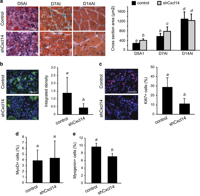
Figure 5.

Cxcl14 knockdown accelerates muscle regeneration post-injury. (a) TA muscles were co-injected with BaCl2 and shRNA viruses, and isolated on days 5, 7 and 14 after injury (AI). Upon cryosection, H&E staining was performed and regenerating myofiber cross-sectional area (CSA) was quantified (n=6 for D5AI, n=8 for D7AI, n=7 for D14AI). (b) TA muscles injected as above were isolated on day 3 AI, cryosectioned and immunostained for Cxcl14 (green) along with DAPI (blue) (n=3). Fluorescence intensity was quantified using ImageJ software. (c–e) TA muscle sections as described in (b) were immunostained for Ki-67 (c), MyoD (d) or myogenin (e), and the percentage of positive cells was quantified (n=5). Scale bars: 50 μm. Paired two-tailed t-test was performed. The data denoted by different letters (a–c) are significantly different from each other (P<0.05). All error bars represent s.d. of independent replicates.
