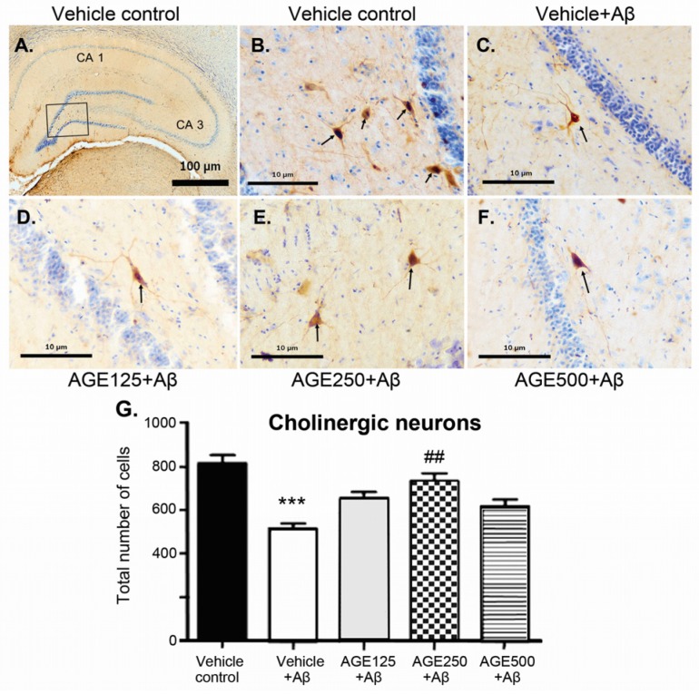
Figure 4.
The neuroprotective effect of AGE (aged garlic extract) on cholinergic neurons in the hippocampal region of Aβ-induced rats. (A–F) represent the photomicrographs of brain section showing the distribution of cholinergic neurons by double staining of Nissl stain with cresyl violet and immunohistochemistry stained with polyclonal ChAT (choline acetyltransferase) antibodies in the vehicle control group (A and B), vehicle + Aβ (C), AGE125 + Aβ (D), AGE250 + Aβ (E) and AGE 500 + Aβ (F). (G) represents the number of ChAT neurons. The cholinergic neurons are indicated with arrows. Data are presented as mean ± S.E.M. (n = 8), *** = significant differences from the vehicle control group at p < 0.001 and ## = significant differences from the vehicle + Aβ group at p < 0.01.

