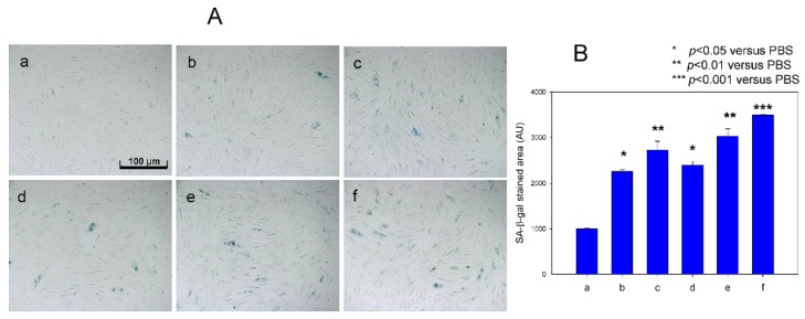
Figure 6.
Induction of cellular senescence in human dermal fibroblasts (HDFs) under presence of Fe2+ by comparison of Senescence-associated β-galactosidase (SA-β-gal) staining. Cells were treated with or without fructose for 15 days (at passage = 13). (A) Cell image was captured using a Nikon Eclipse TE2000 microscope (Tokyo, Japan) at 600× magnification. Photo a, PBS-treated; Photo b, 60 µM Fe2+ (final)-treated; Photo c, 120 µM Fe2+ (final)-treated; Photo d, PBS + fructose (final 5 mM); photo e, fructose (final 5 mM) + 60 µM Fe2+ (final); photo f, fructose (final 5 mM) + 120 µM Fe2+ (final)-treated. (B) Graph shows percentage of SA-β-gal-positive cells per 7.4 mm2 of cell culture area during Fe2+ treatment to human dermal fibroblast (HDF) cells. ***, p < 0.001; **, p < 0.01; *, p < 0.05.

