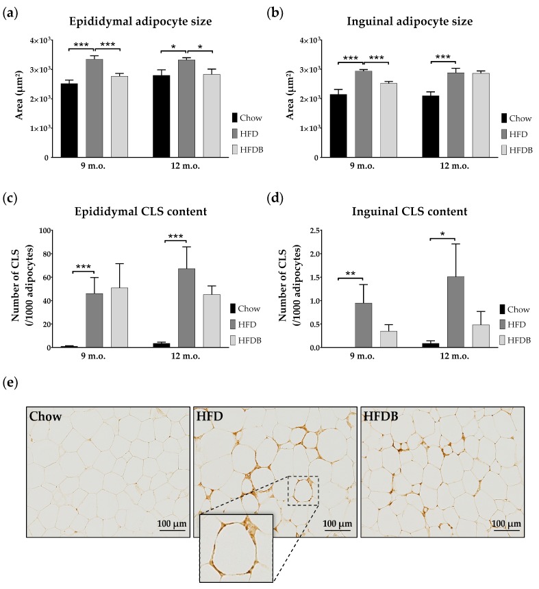
Figure 2.
Adipose tissue cell sizes and macrophage infiltration. Mean adipocyte sizes in epididymal (a) and inguinal (b) adipose tissue depots in both mid- (9 m.o.) and late- (12 m.o.) adult mice. (c,d) Mean number of crown-like structures (CLS) in epididymal (c) and inguinal (d) adipose tissue. (e) Representative photomicrographs of epididymal adipose tissue stained with antibodies against macrophages (MAC-3) in late-adult mice. In addition, a magnification (40×) of a CLS is represented. These examples are comparable to those observed in mid-adulthood. Data are presented as mean ± standard error of mean (SEM). * p < 0.05; ** p < 0.01; *** p ≤ 0.001.

