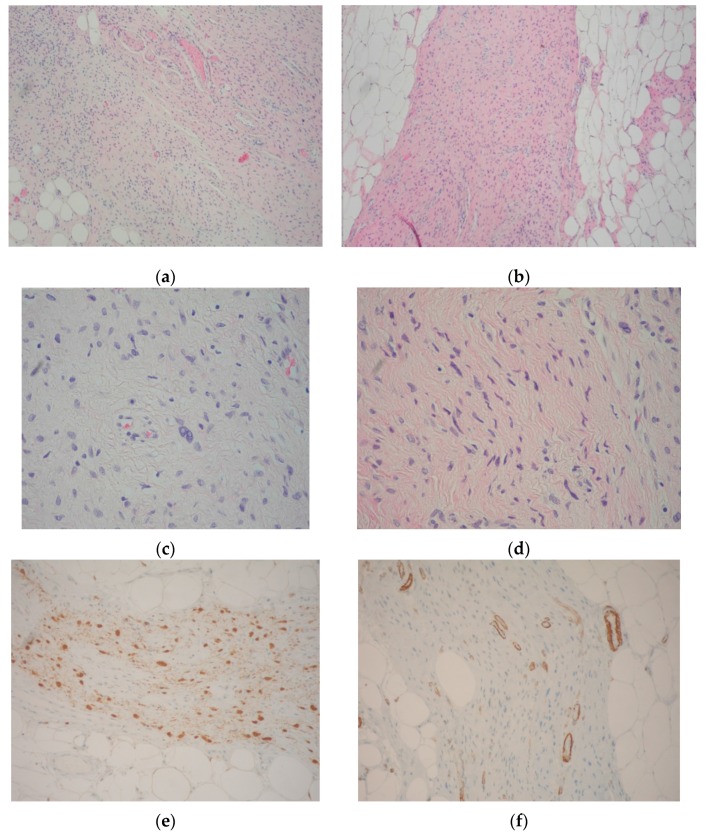
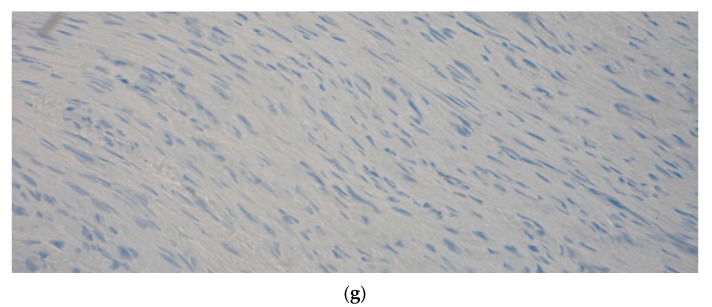
Figure 1.
Neurofibroma resection specimen. (a,b) Spindle “wavy” cells in a matrix of fine fibrillary collagen; neurofibromatous tissue merges with mature fat and ectatic vessels (Hematoxylin-eosin, Magnification 10×); (c,d) Spindle cell nuclei in a fine fibrillary matrix, at a greater magnification (Hematoxylin-Eosin, Magnification 40×); (e) S100 immunohistochemical test confirmed the neurogenic origin of the lesion (Magnification 20×); (f) Actin-hhf35 immunohistochemical test excluded the myogenic origin of the lesion (note that only perivascular splindle smooth cells were reactive) (Magnification 20×); (g) Proliferation index by Ki67 immunohistochemical test was almost negative (Magnification 20×).


