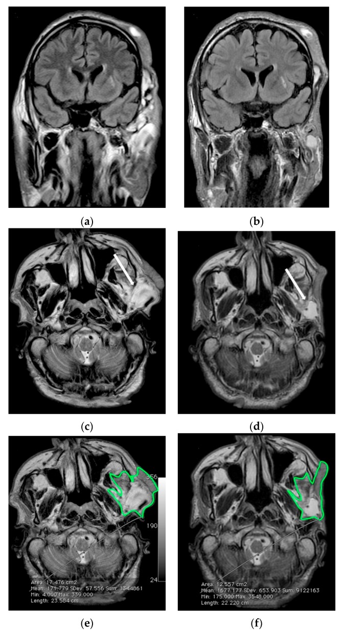
Figure 4.
Serial MR imaging assessment of case 11. (a) Coronal FLAIR-weighted and (c,e) Axial TSE T2-weighted images of the left facial plexiform neurofibroma at baseline; (b) Coronal FLAIR-weighted and (d,f) Axial TSE T2-weighted images after six months of MedDietCurcumin. After six months there was a significant reduction especially in the hyperintense parts of the lesion, as indicated by arrows in (c,d). In (e,f) a green border contours the area used for volume measurement.

