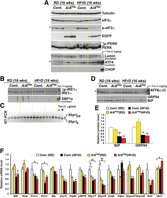
Fig. 4.

High fructose diet deregulates three UPR pathways. a Western blot analysis of PERK pathway proteins in liver lysates from 7-month-old Cont. and A/A Hep mice fed an RD or an HFrD for 16 wks. Nuclear fractions from liver lysates were used to detect ATF4. The volumes of nuclear fractions used were based on nuclear Lamin A/C protein. The liver tissue of a control mouse injected with tunicamycin-(Tm, 1 mg/kg body weight) was used to assess the exact size of ATF4 and CHOP. The * indicates non-specific bands in the Western blot. b Western blot analysis of IRE1α and spliced XBP1 (XBP1s), and RT-PCR analysis of Xbp1 splicing in liver tissues of 7-month-old Cont. and A/A Hep mice fed an RD or an HFrD for 16 wks. The liver tissue of a control mouse injected with Tm was used to assess the exact size of phosphorylated-IRE1α (last lane in the upper panel) and spliced XBP1 proteins (last lane in the lower panel). The same nuclear fractions in Fig. 4a were used to detect nuclear XBP1s. c RT-PCR analysis of Xbp1 mRNA splicing in liver tissues from 7-month-old Cont. and A/A Hep mice fed an RD or an HFrD for 16 wks. Total RNA samples were prepared and RT-PCR analysis was performed with a primer set flanking the intron in unspliced Xbp1 mRNA. PCR products represent unspliced (Xbp1 us) and spliced (Xbp1 s) species. Total RNA samples from mouse embryonic fibroblasts treated with/without Tm (1 μg/ml) were used to indicate the exact sizes of both Xbp1 us and Xbp1 s species. d Western blot analysis of cleaved ATF6α, Grp94 and BiP in liver lysates of 7-month-old Cont. and A/A Hep mice fed an RD or an HFrD for 16 wks. The liver tissue of a control mouse injected with Tm was used to assess the exact size of the N-terminal fragment (ATF6αΔC) of ATF6α (the last lane in the upper panel). The * indicates non-specific bands in the Western blot. e Densitometric quantification of BiP and Grp94 in the lower panel of d). Values were normalized against tubulin levels. Data are means ± SEM (n = 4 mice per group); **p < 0.01 and ***p < 0.001; RD vs HFrD in the same genotype. f Quantitative real-time PCR analysis of the expression of UPR genes in liver tissues from 7-month-old Cont. and A/A Hep mice fed an RD or an HFrD for 16 wks. Data are means ± SEM (n = 5 ~ 6 mice per group); *p < 0.05, **p < 0.01 and ***p < 0.001; RD vs HFrD of the same genotype, # p < 0.05 and ### p < 0.001; Cont. vs A/A Hep , & p < 0.05, && p < 0.01 and &&& p < 0.001; Cont.(RD) vs A/A Hep(HFrD)
