Fig. 2.

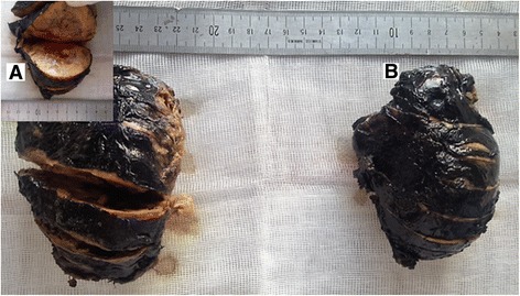
The macroscopic view of the resected adrenal glands. a The right adrenal gland shows a well-encapsulated tumor, the cut surface is yellow-brown, with areas of hemorrhage. b The left resected specimen has quite similar characteristics

The macroscopic view of the resected adrenal glands. a The right adrenal gland shows a well-encapsulated tumor, the cut surface is yellow-brown, with areas of hemorrhage. b The left resected specimen has quite similar characteristics