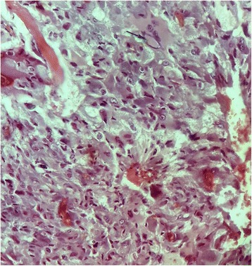
Fig. 4.

The pheochromocytoma component of the tumor shows polygonal, oval-shaped and spindle cells with amphophilic cytoplasm. The nuclei were round to spindle-shaped with inconspicuous nucleoli. A bizarre cell is seen with pseudoinclusion at the top part of the image (black arrow) (Hematoxylin-eosin-safran (HES) ×400)
