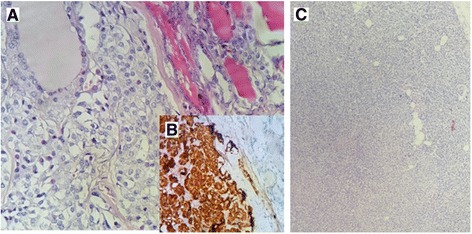
Fig. 7.

The medullary carcinoma of the thyroid shows sheets of polygonal cells with amphophilic cytoplasm and oval nuclei with granular chromatin and inconspicuous nucleoli (a) (HES ×200). These cells are strongly positive for chromogranin (b). The hyperplastic parathyroid shows a significant decrease in adipocytic lobules (c) (Hematoxylin-eosin-safran (HES) ×200)
