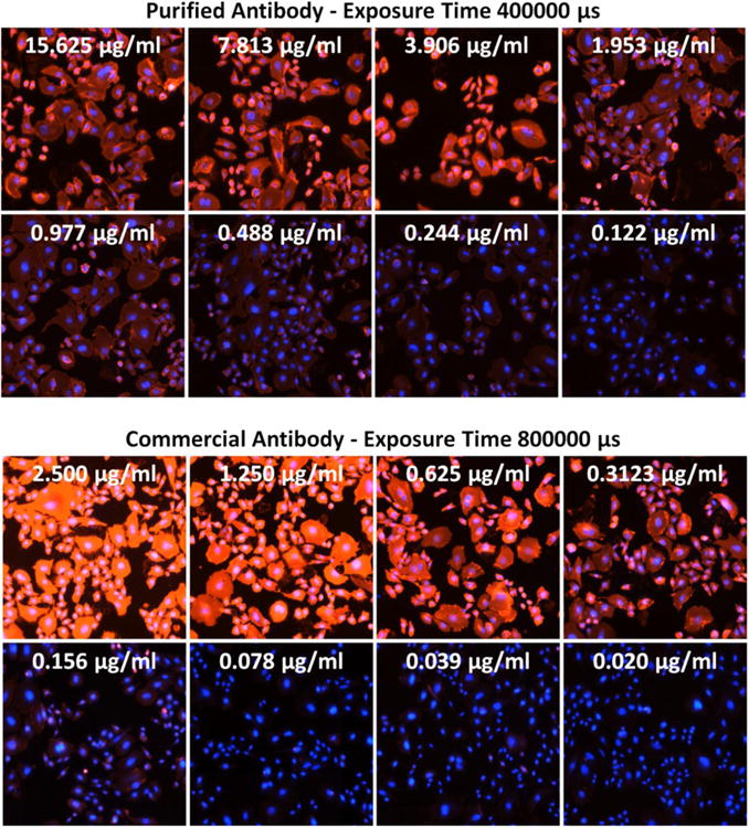
Figure 5.

Overlay of fluorescence images of AF594 (Purified Mab), PE (Commercial Mab), and Hoechst at selected antibody concentrations for purified and commercial antibodies. The exposure times are 400000 and 800000 μs, respectively. The fluorescence intensities were clearly reduced as the antibody concentrations decreased.
