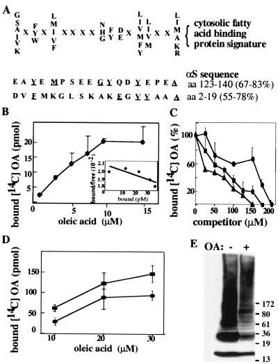
Figure 4.
αS has characteristics of a FABP. (A) The cytosolic FABPs signature. The 18-residue motif comprises nine specific residues and nine undefined residues (represented by X). The homology percentage is calculated either by considering only the specific residues [67% in the amino acid 123–140 stretch (C terminus) and 55% in the amino acid 2–19 stretch (N terminus)] or by including the X residues needed to maintain proper spacing between the specific residues (83% at the C terminus and 78% at the N terminus). Underlined residues are shared with the FABP signature. (B) Purified human αS (5 μM) was incubated with increasing concentrations of 14C OA. Protein-bound OA was separated from free OA by using the Lipidex assay (see Materials and Methods). Data points are means of triplicates ± SD. (Inset) Scatchard plot. Estimated Kd and Bmax values were 12.5 μM and 1, respectively. (C) FA displacement curve: purified human αS (5 μM) was incubated with 10 μM [14C] OA and increasing concentrations of unlabeled OA (●) AA (■) or DHA (▴), as indicated. (D) Aliquots (5 μg) of S370 cytosols from untransfected or wt-αS-transfected MES cells were incubated with increasing concentrations of 14C OA, as in B. Nontransfected cells (●), αS transfected cells (■). (E) Aliquots of S370 (20 μg) were incubated at 65°C overnight without (−) or with (+) free cold OA (5 μM) and Western blotted with LB509.

