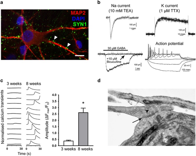
Figure 2.
Physiologically active neurons differentiated from human induced pluripotent stem cell (hiPSC)-derived NPCs. (a) Expression of presynaptic protein Synapsin1 in MAP2-positive neuronal cells. Scale bar=10 μm. (b) Representative graphs of electrophysiological recordings of Na+ and K+ currents, Bicuculline-sensitive GABA-mediated response and evoked action potentials in 8-week-old hiPSC-derived CTL1 neurons. (c) Spontaneous calcium oscillations are more apparent in 8-week-old neurons than in 3-week-old neuronal cultures. Left, representative Ca2+-transients of individual neurons; right, quantification of Ca2+-transients 3 and 8 weeks after terminal differentiation. t-test, *P<0.0001. (d) Presence of synaptic cleft (arrow) and presynaptic vesicles (arrowhead) was verified by electron microscope imaging in 5-week-old hiPSC-derived neurons. Scale bar=500 nm.

