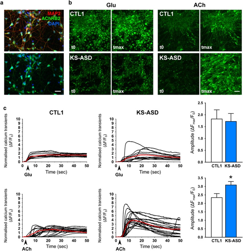
Figure 5.
Increased acetylcholine-evoked calcium signal in neuronal cells differentiated from KS-ASD patient-derived hiPSCs. (a) Expression of Nicotinic Acetylcholine Receptor beta 2 (AChRB2, green) in MAP2-positive 4-week-old hiPSC-derived nerurons. Scale bar=20 μm. (b) Change in fluorescence intensity upon administration of glutamate (Glu) or acetylcholine (ACh) onto 8-week-old control (CTL1) and KS-ASD neurons pre-loaded with Calcium 6-QF dye. Scale bar=50 μm. (c) Representative graphs showing Glu and ACh-evoked Ca-transients in CTL1 and KS-ASD neurons. Right panels, quantification of the Ca2+-responses as the mean of maximal intensity of the responses after treating the cells with Glu (upper) or ACh (lower). t-test, *P<0.05. KS-ASD, Autism spectrum disorder patient with Kleefstra syndrome.

