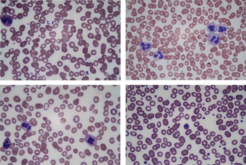
Figure 3.

Peripheral smear examination demonstrating, (a) small scattered platelet clumps; (b) platelet satellitism where platelets are arranged in a rosette-like pattern around the neutrophils; (c) schistocytes, bite cells, and scarcity of platelets, (d) a giant platelet almost the size of a red blood cell.
