
Figure 3.
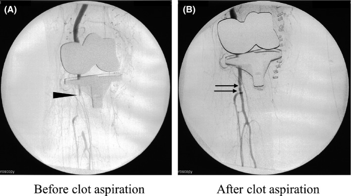
Angiographies of the right knee. (A) Before clot aspiration, the right distal popliteal artery just proximal to the right peroneal‐tibia trunk was occluded (arrow head). (B) After clot aspiration, the peroneal and tibia artery can be confirmed. Stenosis at the distal popliteal artery is still observed at the two portions (arrows).