
Figure 3. Endothelial glycocalyx depth in an individual vessel varies by more than an order of magnitude, according to the imaging and analysis method.
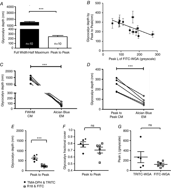
A, microvessels were imaged immediately after perfusion with FITC‐WGA (glycocalyx) and R18 (cell membrane). A line was drawn across the full diameter of the vessel and the depth of the endothelial glycocalyx estimated from: the full width measured at the point of half‐maximal fluorescence intensity of FITC‐WGA (FWHM); and the anatomical distance between peak signal from R18 (cell membrane) and FITC‐WGA (sialic acids within endothelial glycocalyx) (peak to peak). FWHM analysis generated significantly higher values of glycocalyx depth than peak to peak analysis (*** P < 0.001 paired t test). B, the depth of the glycocalyx (peak to peak method) (y‐axis) was inversely related to the peak fluorescence intensity (I f) of FITC‐WGA (x‐axis) (r = −0.50, P < 0.05 Pearson's correlation). C, full‐width half‐maximum (FWHM) measurements of endothelial glycocalyx depth made from confocal microscopy (CM) images were significantly greater than subsequent depth measurements of Alcian Blue‐labelled endothelial glycocalyx imaged with electron microscopy (EM) in the same vessel (*** P < 0.001 paired t test). D, peak to peak measurements of endothelial glycocalyx depth from confocal microscopy (CM) images were also significantly greater than measurements of Alcian Blue‐labelled endothelial glycocalyx depth measurements from electron microscopy (EM) in the same vessel (*** P < 0.001 paired t test). E, glycocalyx depth measurements made in TMA‐DPH and TRITC‐WGA‐labelled vessels by the peak to peak method are significantly greater than peak‐to‐peak‐determined endothelial glycocalyx depth measurements made in R18 and FITC‐WGA‐labelled vessels (n = 6) (*** P < 0.001 unpaired t test). There is no significant difference between fractional glycocalyx cover (F) or peak WGA fluorescence intensity (I f) (G) in TRITC‐WGA‐labelled vessels and FITC‐WGA‐labelled vessels (all non‐significant (ns), P > 0.05 unpaired t test).