
Figure 5. Disruption of sialic acids reduces endothelial glycocalyx depth and coverage.
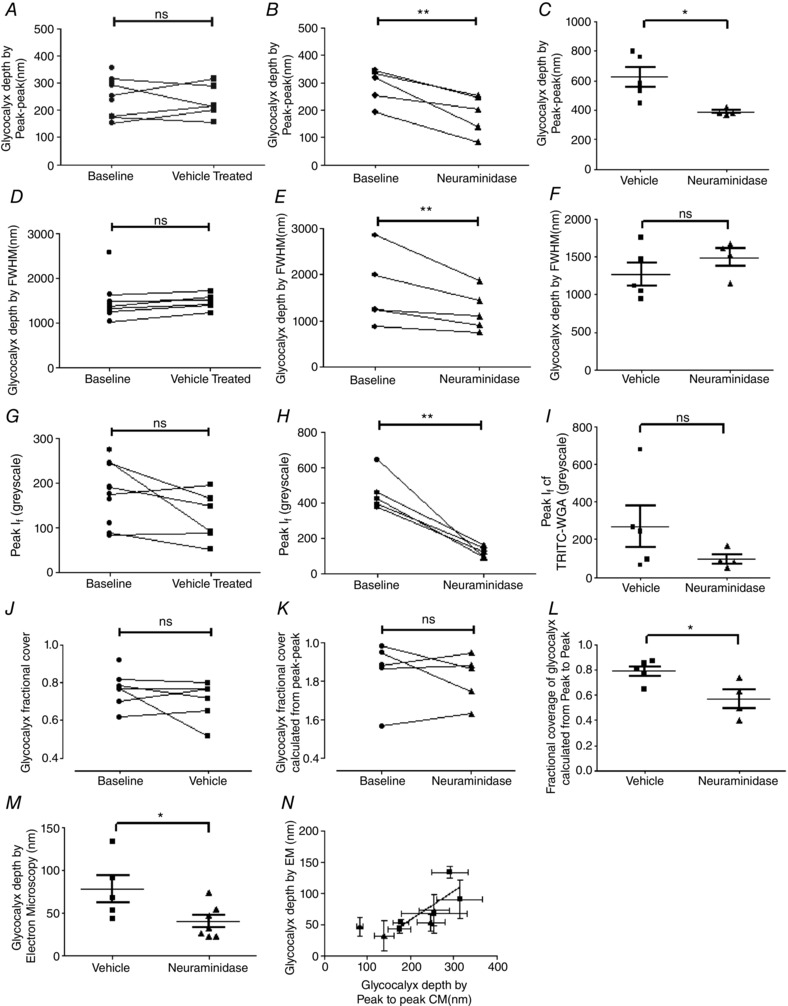
Endothelial glycocalyx parameters were determined in vessels with sialic acid disruption achieved by 20 min perfusion with 40 mg ml−1 BSA supplemented with 2 U ml−1 neuraminidase (filled triangles), or in control vessels perfused with 40 mg ml−1 BSA vehicle alone (baseline: filled circles; vehicle perfusion; filled squares). Glycocalyx depth determined using the peak to peak method of analysis was no different before and after perfusion with vehicle (A), but was significantly reduced after neuramindase perfusion (B), (ns: P > 0.05; ** P < 0.01, two‐way ANOVA with Bonferroni post hoc analysis). This neuraminidase‐induced reduction in peak‐to‐peak‐determined measurements of endothelial glycocalyx depth was reproduced in unpaired experiments (C), in which endothelial glycocalyx depth was determined once only, after earlier measurements of solute permeability in the same vessel during perfusion with either 40 mg ml−1 BSA vehicle alone (filled squares) or 40 mg ml−1 BSA supplemented with 2 U ml−1 neuraminidase (filled triangles) (* P < 0.05, unpaired t test). Likewise, glycocalyx depth determined with the full‐width half‐maximum (FWHM) method was unaltered by perfusion with vehicle solution (D), but was significantly reduced after neuraminidase perfusion (E) (ns: P > 0.05; ** P < 0.01, two‐way ANOVA with Bonferroni post hoc analysis). However, in separate measurements in which endothelial glycocalyx depth was measured only once by the FWHM after initial solute permeability measurements during perfusion with either 40 mg ml−1 BSA vehicle alone (filled squares) or 40 mg ml−1 BSA supplemented with 2 U ml−1 neuraminidase (filled triangles), no significant difference was determined (ns, P > 0.05, unpaired t test) (F). Identical results (as for FWHM) were also obtained when determining the peak I f of WGA‐labelled endothelial glycocalyx in paired (G and H; ns: P > 0.05; ** P < 0.01, two‐way ANOVA with Bonferroni post hoc analysis) and unpaired (I; ns, P > 0.05, unpaired t test) experiments. The fractional coverage of the vessel wall with endothelial glycocalyx resolvable with the peak to peak method of analysis was no different before and after perfusion with either vehicle or neuraminidase (J and K; ns: P > 0.05, two‐way ANOVA with Bonferroni post hoc analysis), but a significant reduction was observed after neuraminidase perfusion in unpaired experiments (L; * P < 0.05, unpaired t test). Endothelial glycocalyx depth measured in electron micrographs was significantly lower in neuramindase‐perfused vessels, as compared with vehicle‐perfused vessels (* P < 0.05, unpaired t test) (M). There was a significant positive correlation between the depth of endothelial glycocalyx determined in electron microscopy (EM) images with the depth of endothelial glycocalyx determined by the peak to peak analysis method in confocal microscopy (CM) images in the same vessel (N; r = 0.75, P < 0.05 Pearson correlation).