Fig. 6.
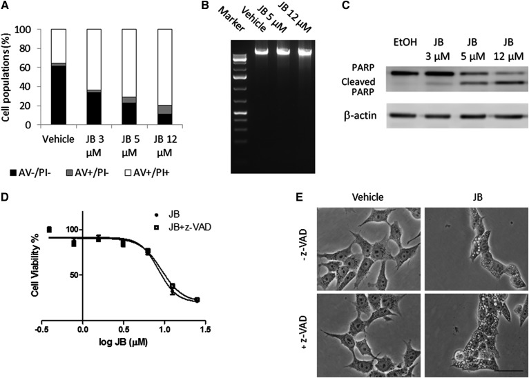
JB induces nonapoptotic cell vacuolation in gastric cancer cells. A: Cells were incubated for 16 h with increasing concentrations of JB or ethanol and stained with annexin V (AV)/PI and fluorescence was analyzed by flow cytometry. The percentage of cell populations stained with AV, PI, or both are shown. Results are representative of a typical experiment in triplicate that gave similar results (n = 3). B: HGC-27 cells were treated with JB (5 or 12 μM) or ethanol (0.12%) as a control for 16 h. After extraction, DNA was run on agarose gel containing SYBR green. One hundred kilobases of DNA standard were used as a marker. The image represents a typical experiment repeated two times that gave similar results. C: HGC-27 cells were treated with JB (3, 5, and 12 μM) or ethanol as a control for 16 h. PARP and cleaved PARP were detected by Western blot. β-Actin was used as a loading control. The results are representative of four different experiments. D: JB cytotoxicity was assessed by MTT after incubating HGC-27 cells with JB for 16 h in the absence (JB) or presence (JB + z-VAD) of the pan-caspase inhibitor, z-VAD (20 μM). The results are the mean ± SD of two independent experiments repeated in triplicate. E: Phase-contrast images of cells incubated with JB (5 μM) or ethanol for 16 h in the presence of z-VAD (20 μM) (+z-VAD) or DMSO (−z-VAD). The images are representative of the observed phenotypes (n = 2).

