Fig. 7.
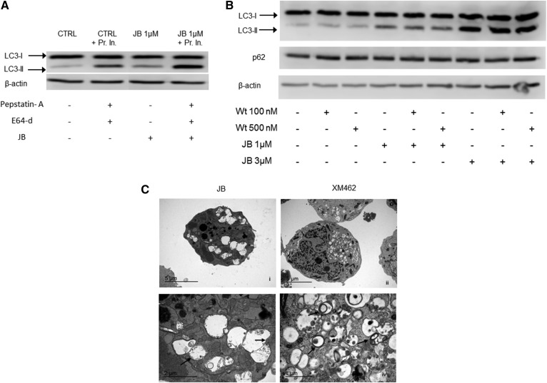
JB causes autophagy-independent LC3-II accumulation. A: HGC-27 cells were treated with JB (1 μM) (or ethanol) for 16 h with or without protease inhibitors [pepstatin-A (8 μM) and E64-d (30 μM)] that were added 2 h before the treatment. B: Cells were incubated with wortmannin (Wt) (100 or 500 nM) (or DMSO) for 1 h and then with JB (1 or 3 μM) (or ethanol) for 16 h. LC3-I and LC3-II levels or p62 were detected by Western blot. β-Actin was used as a loading control. Results are representative of two to three different experiments. C: Transmission electron micrographs of HGC-27 cells treated with JB (5 μM) (i) or XM462 8 μM (ii) for 16 h. Images (iii) and (iv) represent magnifications of (i) and (ii), respectively. Arrows indicate single (ii) or double (iv) membrane vesicles. The experiment was run in triplicate; images are representative of the observed phenotypes.

