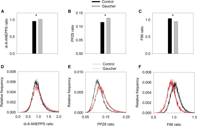
Fig. 4.
Effect of sphingolipid accumulation in a model of Gaucher’s disease on the dipole potential. THP-1 monocytes were differentiated to macrophages with PMA in the absence (control) and presence (Gaucher) of CBE, followed by labeling them with three different dipole potential-sensitive dyes: di-8-ANEPPS (A, D), PPZ8 (B, E), and F66 (C, F). The intensity ratios were calculated in 20–30 cells, and their means (±SEM) are displayed in the bar graphs (A–C). Representative histograms showing the distribution of pixelwise fluorescence ratios are displayed in (D–F). *P < 0.05, significant difference found between the intensity ratios in control and Gaucher-type cells by two-way ANOVA, followed by Tukey’s HSD test.

