
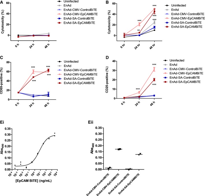
Figure EV3. Cytotoxicity and T‐cell activation by EnAd expressing EpCAM BiTE in DLD cells.
-
A, BCytotoxicity for infected DLD cells in the absence (A) or presence of T cells (B). DLD cells were infected and co‐cultured with T cells, and cytotoxicity was measured by LDH release at the specified time points.
-
C, DT cells from (B) were harvested and stained for activation markers CD69 (C) or CD25 (D) and analysed via flow cytometry.
-
EQuantification of EpCAM BiTE produced from DLD cells infected with recombinant viruses. Standard curve of LDH released (Abs) of DLD cells in co‐culture with CD3+ cells and serial dilutions of a known quantity of recombinant EpCAM BiTE (Ei). In parallel, co‐cultures were incubated with diluted supernatants (10,000‐fold) from 3‐day infected DLD cells (Eii). Standard curve allowed the approximate determination of EpCAM BiTE produced at 165 and 50 μg per million DLD cells for EnAd‐CMV‐EpCAMBiTE and EnAd‐SA‐EpCAMBiTE, respectively.