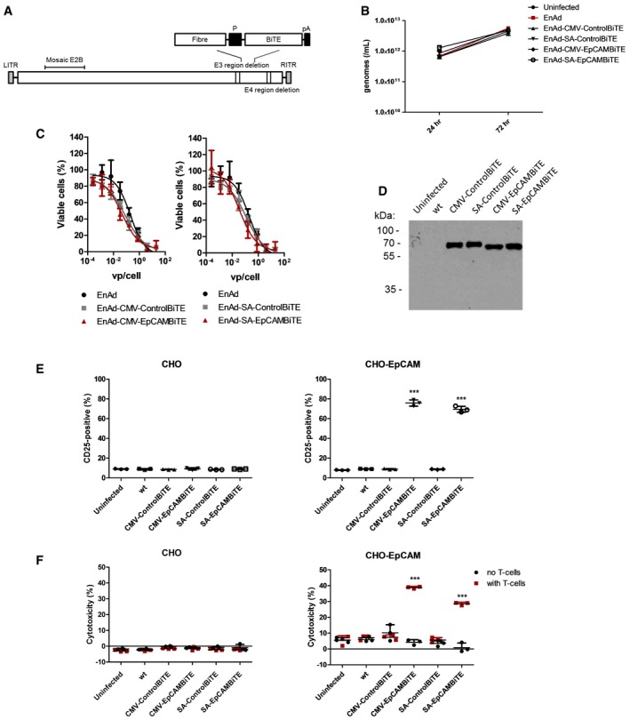
Schematic representation of EnAdenotucirev engineered to express the BiTE transgenes downstream of a CMV promoter or splice acceptor site (P) followed by a polyadenylation sequence (pA).
DLD cells were infected with parental EnAd or recombinant virus (100 vp/cell; 2.5e7 vp/ml) and wells harvested at 24 or 72 h. Replication was assessed by measuring genomes using qPCR against viral hexon.
Cytotoxicity of DLD cells infected with EnAd or recombinant virus at increasing concentrations of virus. Cytotoxicity was measured by MTS assay after 5 days of infection.
Supernatants from day 3 uninfected or virus‐infected HEK293A cells were assessed for transgene expression by immunoblot analysis and probed with an anti‐His antibody.
Induction of activation marker CD25 of CD3‐positive T cells cultured with CHO or CHO‐EpCAM (E:T 5:1) and diluted HEK293A supernatants from (D). Activation was measured by surface expression of CD25 by flow cytometry.
Cytotoxicity of CHO or CHO‐EpCAM cells incubated with HEK293A supernatants from (D) alone or in co‐culture with CD3‐purified PBMC (E:T 5:1). HEK293A supernatants were diluted 300‐fold. Cytotoxicity was assessed by LDH released into the supernatant after 24‐h incubation.
Data information: Each condition was measured in biological triplicate and represented as mean ± SD. Significance was assessed using a one‐way ANOVA test with Tukey's
post hoc analysis with each condition compared to untreated, ***
P < 0.001.
Source data are available online for this figure.