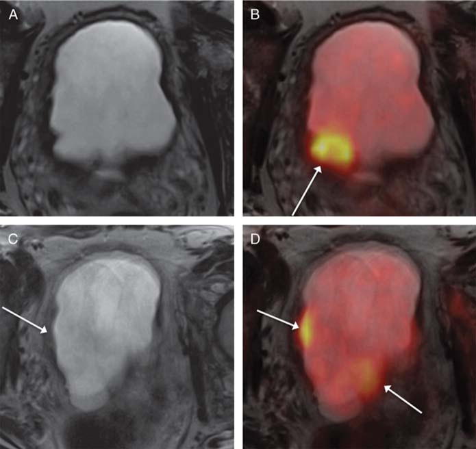
FIGURE 1.

A 68-year-old man with muscle-invasive high-grade bladder cancer on prior biopsy, undergoing simultaneous 18F-FDG PET/MRI. A and B, Axial T2-weighted images show regions of mild nonspecific mural thickening (eg, arrow, B) that was considered equivocal for presence of tumor. C and D, Fused PET/MR images show multiple focal regions of increased metabolic activity (arrows, C and D), which increased confidence for the presence of bladder tumor. Subsequent radical cystectomy demonstrated multifocal tumor.
