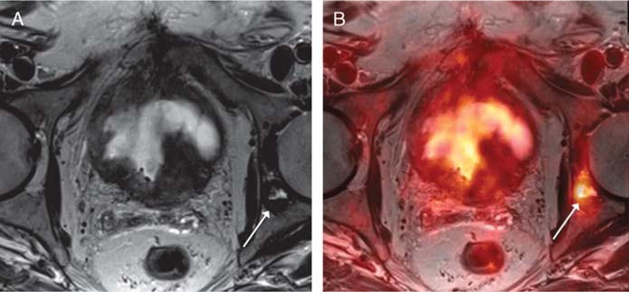
FIGURE 7.

An 82-year-old man with prior biopsy showing high-grade non–muscle-invasive bladder cancer, undergoing simultaneous 18F-FDG PET/MRI. A, Axial T2-weighted image shows a left acetabular lesion (arrow) that was considered possibly degenerative, given its proximity to the hip joint, and equivocal for osseous metastasis. B, Fused PET/MR image shows corresponding marked increased metabolic activity (arrow), raising suspicion that the lesion represents an osseous metastases. Subsequent bone biopsy demonstrated metastatic urothelial carcinoma.
