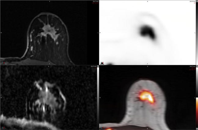
Fig. 1.

54-year-old woman with newly diagnosed left breast cancer. FDG PET-MR imaging performed in the prone position (reoriented) with a dedicated breast coil demonstrates intense FDG uptake in primary tumor on PET and PET-MR images (top right and bottom right). ADC map MR image (bottom left) demonstrates heterogeneous signal intensity with areas of low ADC (grey-black regions) within tumor due to high cellularity. Postcontrast T1-weighted MR image (top left) demonstrates heterogeneous enhancement within the mass. Prone PET-MR imaging with dedicated breast coil facilitates multiparametric quantitative analysis of primary breast tumors. (Courtesy of Dr Amy Melsaether, NYU Langone Medical Center, New York, NY.)
