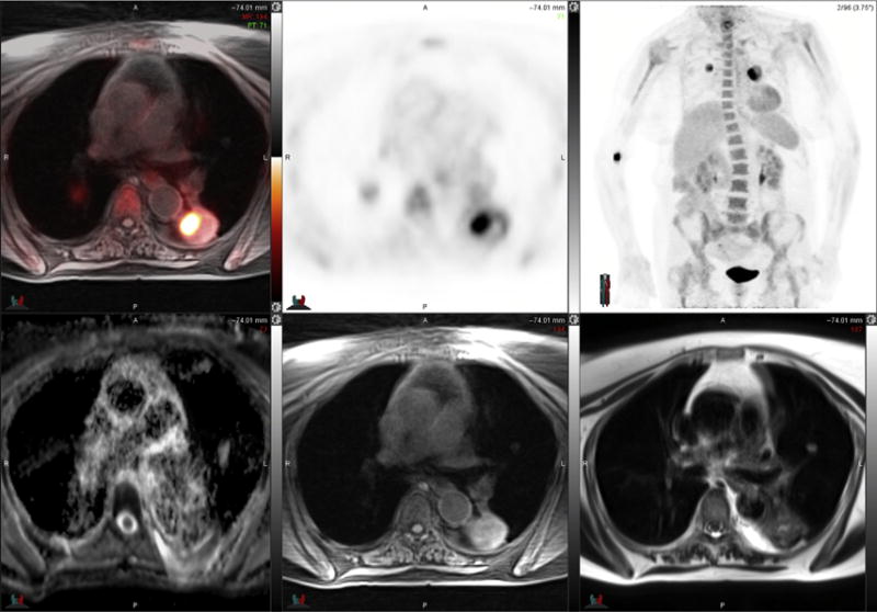
Fig. 4.

78-year-old woman with bilateral lung cancers. FDG PET-MR image (top left) and PET images (top middle and top right) demonstrate peripheral intense radiotracer uptake in left lung tumor that is most intense medially. ADC map MR image (bottom left) is degraded by motion artifact, a significant challenge for lung nodule imaging. T1-weighted fat-suppressed MR image (bottom middle) and T2-weighted MR image (bottom right) demonstrate heterogeneous signal intensity in the left lower lobe mass.
