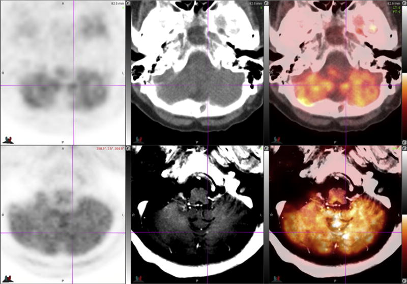
Fig. 6.

70-year-old woman with NSCLC. FDG PET-MR images (bottom row) identify an enhancing tiny new left cerebellar brain metastasis seen only on postcontrast T1-weighted MR image (bottom middle, see crosshair) and PET-MR image (bottom right, see crosshair). Note that the lesion is not visible on PET images (left column), low-dose unenhanced CT image (top middle), or on PET-CT image (top right).
