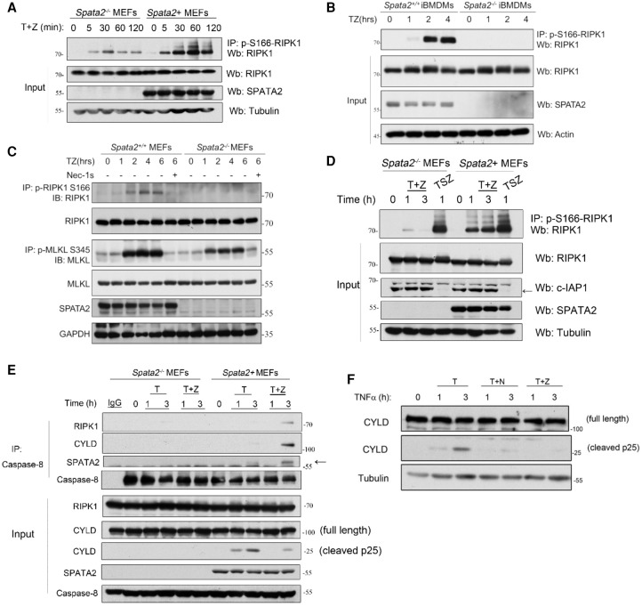
Figure 2.
SPATA2 promotes the activation of the RIPK1 kinase and the formation of complex IIb. (A) Immortalized Spata2−/− MEFs and Spata2+ MEFs were treated with 100 ng/mL human TNF and 50 µM zVAD.fmk for the indicated periods of time. The cell lysates were analyzed by immunoprecipitation using p-S166 RIPK1 antibody followed by Western blotting using the indicated antibodies. (B) Spata2+/+ iBMDMs and Spata2−/− iBMDMs were treated with 100 ng/mL human TNFα plus 50 µM zVAD.fmk for the indicated periods of time. The cell lysates were subjected to immunoprecipitation using anti-p-S166-RIPK1 antibody, and the immunocomplexes were analyzed by Western blotting. (C) Immortalized Spata2+/+ MEFs and Spata2−/− MEFs were pretreated with 20 µM Nec-1s for 30 min and then treated with 50 ng/mL human TNFα and 50 µM zVAD.fmk for the indicated periods of time. The cell lysates were analyzed by immunoprecipitation using anti-p-S166 RIPK1 or p-S345 MLKL antibody as indicated. The immunocomplexes or cell lysates were analyzed by Western blotting using the indicated antibodies. (D) Immortalized Spata2−/− MEFs and Spata2+ MEFs were pretreated with or without 100 nM SM-164 for 30 min and then treated with 100 ng/mL human TNFα for the indicated periods of time. The cell lysates were analyzed by immunoprecipitation using anti-p-S166 and analyzed by Western blotting. (E) Immortalized Spata2−/− MEFs and Spata2+ MEFs were treated with 100 ng/mL human TNFα and 20 µM zVAD.fmk for the indicated periods of time. The cell lysates were analyzed by immunoprecipitation using anti-caspase-8 followed by Western blotting using the indicated antibodies. (F) MEFs were pretreated with or without 20 µM Nec-1s for 10 min and then treated with 100 ng/mL human TNFα and 50 µM zVAD.fmk for the indicated periods of time. The cell lysates were analyzed by Western blotting using the indicated antibodies. GAPDH and Tubulin were used as loading controls.

