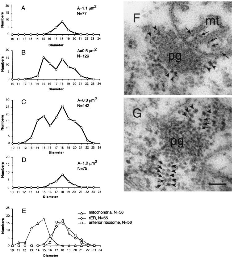
Figure 3.
Two types of ribosomes in the polar-granule polysomes. (A–D) The average number of ribosomes in a unit area (0.3 μm2) is plotted against its diameter. Diameters of the ribosomes around polar granules were measured in stage-14 oocytes (A), stage-1 embryos (B), stage-2 embryos (C), and in pole cells of stage-4 embryos (D). In A–D, total area examined (A) and total number of the ribosomes counted (N) are shown. (E) The number of ribosomes within mitochondria, in the anterior region and on the surface of rough endoplasmic reticulum, are plotted against their diameters. (F and G) Electron micrographs of sections through polar granules. (F) In stage-1 embryos, polar granules are intimately associated with mitochondria, and no ribosomes were found at the boundary between these organelles. Arrows point to ribosomes within a mitochondrion. (G) In stage-2 embryos, the smaller ribosomes (arrows) are integrated into the polar granule-polysomes. Arrowheads (F and G) point to the larger ribosomes. pg, polar granules; mt, mitochondria. (Scale bar = 0.1 μm.)

