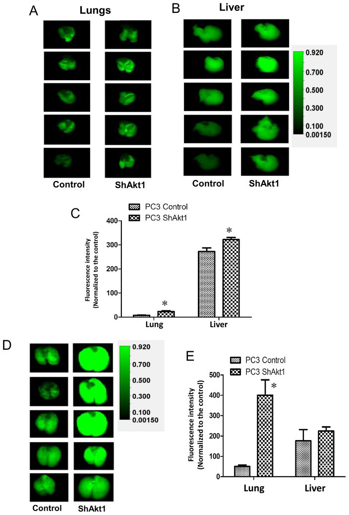
Figure 4. Akt1 loss in human PC3 prostate cancer cells promotes lung metastasis.
(A) Representative images of IRDye 800CW 2-deoxy glucose loaded lungs and (B) livers from athymic nude mice pre-administered (via tail-vein; 3 days ago) with Control ShRNA (Control) or Akt1 ShRNA (ShAkt1) expressing human PC3 cells. (C) Bar graph showing the quantified fluorescent intensity of uptaken IRDye 800CW 2-deoxy glucose by the lungs and liver in athymic nude mice pre-administered with ShControl or ShAkt1 expressing human PC3 cells (n=8). (D) Representative images of IRDye 800CW 2-deoxy glucose loaded lungs from athymic nude mice pre-administered (via tail-vein; 2 weeks ago) with ShControl or ShAkt1 expressing human PC3 cells. (E) Bar graph showing the quantified fluorescent intensity of uptaken IRDye 800CW 2-deoxy glucose by the lungs and liver in athymic nude mice pre-administered (via tail-vein; 2 weeks ago) with ShControl or ShAkt1 expressing human PC3 cells (n=8). Data presented as Mean ± SD; *P < 0.05.

