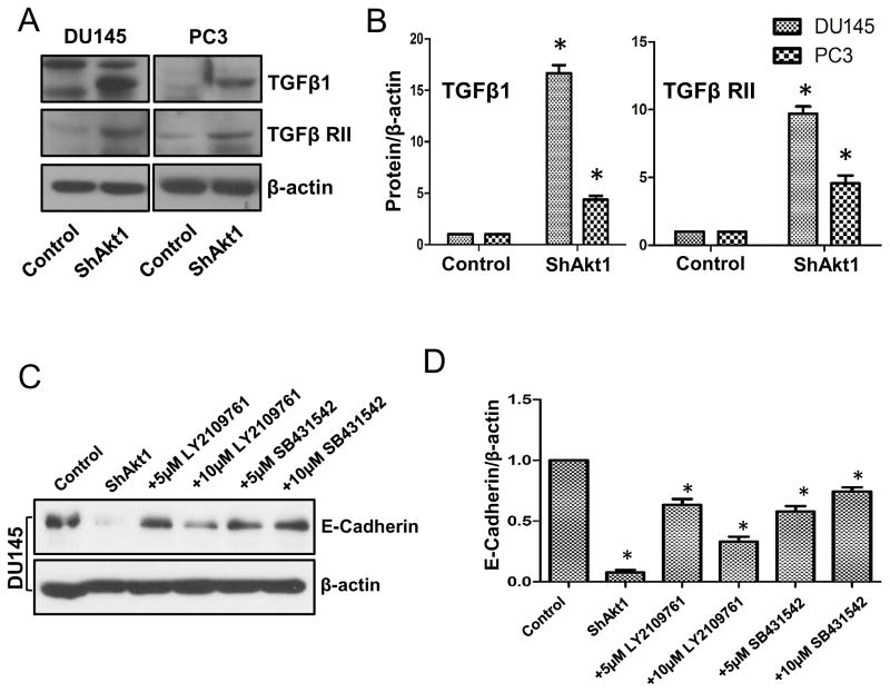
Figure 7. Akt1 suppression in human prostate cancer cells result in increased expression of EMT-inducing growth factor TGFβ1 and its receptor TGFβ RII.
(A) Representative Western blot images of control and Akt1 knockdown PC3 and DU145 cell lysates showing changes in the expression levels of TGFβ1 and its receptor TGFβ RII compared to respective control cell lysates. (B) Bar graphs showing average fold-changes in the expression levels of TGFβ1 and its receptor TGFβ RII in Akt1 knockdown PC3 and DU145 cell lysates compared to respective control cell lysates (n=4). (C) Representative Western blot images of control and Akt1 knockdown DU145 cell lysates showing changes in the expression levels of epithelial marker E-cadherin in the presence and absence of DMSO (control), and various doses of TGFβ receptor inhibitors LY2109761 and SB431542 compared to control cell lysates. (D) Bar graphs showing average fold-changes in the expression levels of E-cadherin in Akt1 knockdown DU145 cell lysates treated in the presence and absence of DMSO (control), and various doses of TGFβ receptor inhibitors LY2109761 and SB431542 compared to control cell lysates (n=4). Data presented as Mean ± SD; *P < 0.05.

