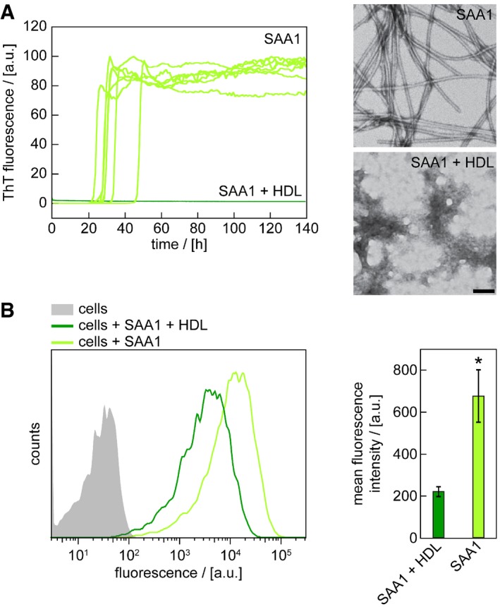
Figure 6. HDL‐binding sequesters SAA1 protein and prevents fibril formation.

- Time‐dependent ThT fluorescence measurements of 50 μM SAA1 in the presence or absence of HDL. Seven replicates per condition are shown. The lag time is 31.5 ± 6.8 h in the absence of HDL. TEM images were taken from samples after 140 h. Scale bar: 100 nm.
- Flow cytometric analysis of cells incubated with 0.5 mg/ml SAA1, 0.01 mg/ml SAA1‐AF488 and HDL as indicated for 5 h (n = 3). Data are presented as mean ± SD. *P < 0.05 (Student's t‐test).
