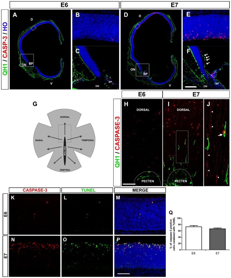
Fig 1. The onset of microglial entry into the embryonic quail retina coincides with an increase in the death of retinal cells.
(A, D) Confocal images of QH1 (green) and anti-active caspase-3 (red) double-immunostained cross sections from quail embryo eyes at 6 (E6, A) and 7 (E7, D) days of incubation. Cell nuclei are stained with Hoechst 33342 (blue). D: dorsal; V: ventral; BP: base of the pecten; ON: optic nerve. (B, E) Higher magnifications of the upper boxed areas in A and D, respectively. Active caspase-3-expressing cells (dying cells) are almost non-existent in the E6 retina (B) and are markedly increased in the dorsal region of the E7 retina (E). (C, F) Higher magnifications of the lower boxed areas in A and D, respectively. No QH1-positive microglial cells are seen in the retina at E6 (C), whereas some (arrows) are migrating within the retina at E7 in the vitreal part of the region dorsal to the ON and BP (F). Scale bar in F: 725 μm for A and D; 50 μm for B and E; 160 μm for C and F. (G) Schematic drawing representing a whole-mounted quail embryo retina, with the base of the pecten (black area) in a central location, from which microglia migrate in a central-to-peripheral direction (arrows) from 7 days of incubation (E7) onwards. The area delimited with dashed lines represents the retinal zone immediately dorsal to the pecten, shown in H and I. (H-J) Confocal images of the region dorsal to the base of the pecten (BP) of QH1 (green) and anti-active caspase-3 (red) double-immunostained whole-mounted quail embryo retinas at E6 (H) and E7 (I). No microglial cells are present within the E6 retina, in which caspase-3-positive dying cells are almost absent (H). By contrast, at E7, some elongated microglial cells enter the retina from the BP and are directed toward a retinal area with numerous caspase-3-positive cells (I). Higher magnification of the boxed area in I is displayed in J, showing the close contact between a migrating microglial cell and the soma of a caspase-3-positive cell (arrow). Another microglial cell is in close contact with long thin caspase-3-positive processes (arrowheads) that appear to be axons from dying cells. Scale bar in H: 100 μm for H and I; 37 μm for J. (K-P) Anti-active caspase-3 (red) and TUNEL (green) double-labeled cross-sections from E6 (K-M) and E7 (N-P) retinas. The co-localization of anti-active caspase-3 immunolabeling and TUNEL staining in numerous cells demonstrates that they are apoptotic cells. Scale bar in P: 50 μm for K-P. (Q) Percentage of active caspase-3 positive cells colabeled with TUNEL in E6 (white bar) and E7 (grey bar) retinas. Data are expressed as means ± SEM (n = 3 for each age). Despite the much higher presence of active caspase-3-positive cells in E7 than in E6, the percentage of caspase-3/TUNEL-positive cells is very similar in each developmental stage.

