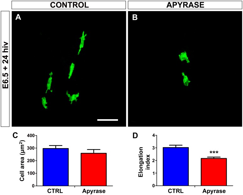
Fig 5. In vitro treatment with apyrase induces rounding of microglial cells in quail embryo retina explants.
(A, B) QH1-positive microglial cells (green) in retina explants from a quail embryo at 6.5 days of incubation cultured for 24 hours in vitro (E6.5+24hiv) in apyrase-free (CONTROL, A) or apyrase-containing (APYRASE, B) medium. Microglial cells in the apyrase-treated explant are more rounded and less elongated (B) than those in the control explant, where they show the typical morphology of cells migrating tangentially in the retina, with an elongated cell body and polarized processes (A). Scale bar: 50 μm. (C, D) Morphometric analysis of cell area (C) and elongation index (D) for microglial cells in non-treated (CTRL, blue bars) and apyrase-treated (Apyrase, red bars) E6.5+24hiv retina explants. Data are expressed as means ± SEM (n = 24). Asterisks depict significant differences (***P<0.001, Student´s t-test). Although the cell area does not significantly differ between apyrase-treated and non-treated explants (C), the elongation index is significantly lower in apyrase-treated explants than in non-treated controls (D).

