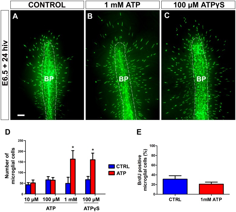
Fig 9. In vitro treatment with 1 mM ATP or non-hidrolyzable ATP (ATPγS) stimulates entry of microglial cells into the embryonic quail retina.
(A-C) QH1-immunolabeled (green) whole-mounted retina explants from quail embryos at 6.5 days of incubation cultured for 24 hours in vitro (E6.5+24hiv) in ATP-free medium (CONTROL, A) or in medium containing 1mM ATP (B) or 100 μM ATPγS (C). QH1-labeled microglial cells appear to be more abundant in ATP- and ATPγS-treated retina explants than in the non-treated explant. BP: base of the pecten (delimited with a dashed line). Scale bar: 100 μm. (D) Number of microglial cells migrating within the retina in explants treated with different concentrations of ATP (10 μM, 100 μM, and 1 mM) or 100 μM ATPγS (red bars) and in their respective non-treated controls (blue bars). Data are expressed as means ± SEM (n = 10 for each). Asterisks depict significant differences (*P<0.05, Student´s t-test). Treatments with 1 mM ATP and 100 μM ATPγS significantly increase the number of microglial cells in E6.5+24hiv retina explants. In contrast, no significant differences are observed between explants treated with low ATP concentrations (10 μM and 100 μM) and their non-treated controls. (E) Percentages of BrdU-positive microglial cells in retina explants treated with 1 mM ATP (red bar) and non-treated controls (blue bar). Data are expressed as means ± SEM (n = 8). No significant differences are detected between ATP-treated and non-treated explants.

