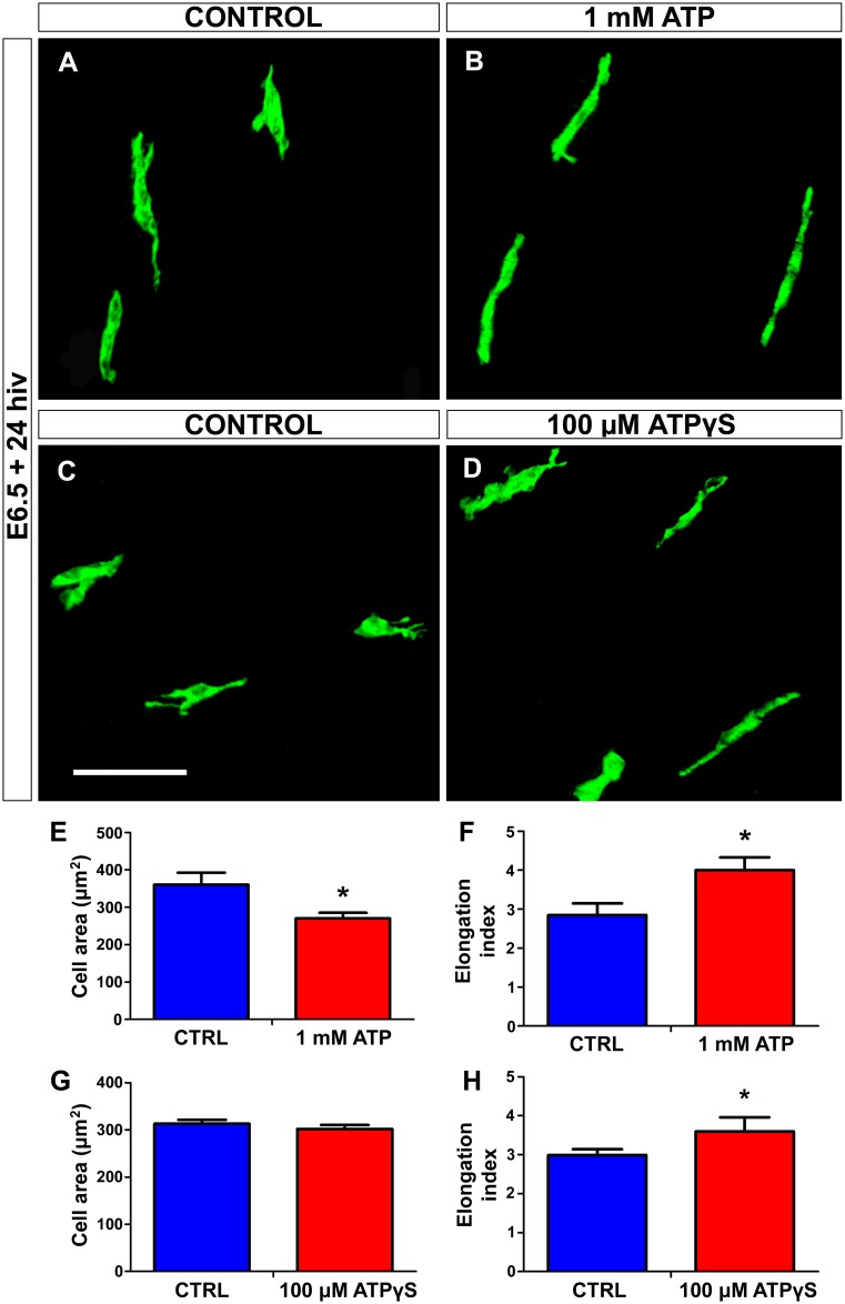
Fig 10. In vitro treatment with ATP or non-hydrolyzable ATP (ATPγS) induces elongation of microglial cells in quail embryo retina explants.
(A-D) QH1-immunolabeled microglial cells (green) in retina explants from quail embryos at 6.5 days of incubation cultured for 24 hours in vitro (E6.5+24hiv) in ATP-free (CONTROL, A), 1 mM ATP-containing (1 mM ATP, B), ATPγS-free (CONTROL, C), or 100 μM ATPγS-containing (D) medium. Microglial cells appear more elongated in ATP- and ATPγS-treated explants than in their respective control explants, which is compatible with a greater migratory activity of microglial cells in nucleotide-treated explants. Scale bar: 50 μm. (E-H) Morphometric analysis of cell area (E and G) and elongation index (F and H) for microglial cells in non-treated (CTRL, blue bars), 1 mM ATP-treated (red bars), and 100 μM ATPγS-treated (red bars) E6.5+24hiv retina explants. Data are expressed as means ± SEM (n = 30 for each). Asterisks depict significant differences (*P<0.05, Student´s t-test). The elongation index is significantly higher in 1 mM ATP- and 100 μM ATPγS-treated explants than in their respective non-treated controls.

