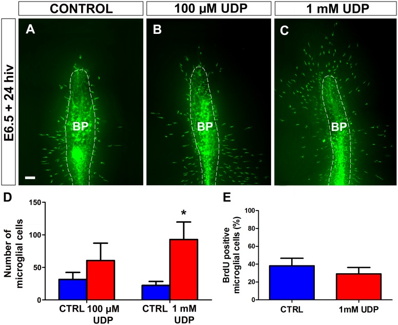
Fig 11. In vitro treatment with UDP stimulates entry of microglial cells into the embryonic quail retina.
(A-C) QH1-immunolabeled (green) whole-mounted retina explants from quail embryos at 6.5 days of incubation cultured for 24 hours in vitro (E6.5+24hiv) in UDP-free medium (CONTROL, A) or in media containing 100 μM UDP (B) or 1 mM UDP (C). QH1-labeled microglial cells appear more abundant in both UDP-treated retina explants than in the non-treated explant. BP: base of the pecten (delimited with a dashed line). Scale bar: 100 μm. (D) Number of microglial cells migrating within the retina in 100 μM UDP- and 1mM UDP-treated E6.5+24hiv retina explants (red bars) and in their respective non-treated controls (blue bars). Data are expressed as means ± SEM (n = 10 for each). Asterisk depicts a significant difference (*P<0.05, Student´s t-test). The number of microglial cells entering the retina increases after UDP treatment at both concentrations, but the increase only reaches significance at the higher concentration (1 mM). (E) Percentages of BrdU-positive microglial cells in retina explants treated with 1 mM UDP (red bar) and non-treated controls (blue bar). Data are expressed as means ± SEM (n = 8). No significant differences are observed between UDP-treated and non-treated explants.

