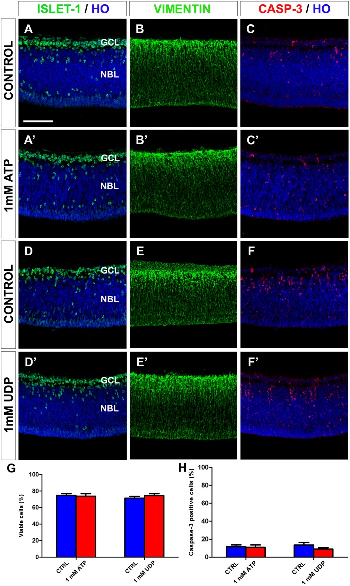
Fig 13. In vitro treatment with ATP or UDP does not alter cell viability or the distribution patterns of active caspase-3-, islet-1-, or vimentin-positive cells in quail embryo retina explants.
(A-F) Single anti-islet-1 (A, A’, D, D’), anti-vimentin (B, B’, E, E’) and anti-active caspase-3 (C, C’, F, F’) immunostained cross-sections of retina explants from quail embryos at 6.5 days of incubation cultured for 24 hours in vitro in the presence of 1 mM ATP (A’, B’, C’) or 1 mM UDP (D’, E’, F’). Cross sections of ATP-free and UDP-free explants (CONTROL) are displayed in A, B, C and D, E, F, respectively. Cell nuclei are stained with Hoechst 33342 (HO, blue). Islet-1-positive neurons are mainly located in the ganglion cell layer (GCL). Vimentin-positive cells are distributed throughout the retina thickness, with higher expression in the GCL. Caspase-3-positive dying cells are mainly located in the GCL and the inner half of the neuroblastic layer (NBL). The retina cytoarchitecture and caspase-3 expression pattern remain unaltered after ATP or UDP treatment. Scale bar: 50 μm. (G, H) Percentages of viable cells (G) and caspase-3-positive cells (H), as determined by flow cytometry, in ATP- or UDP-treated explants (red bars) and their respective non-treated controls (blue bars). Data are expressed as means ± SEM (n = 6 for each). No significant differences in cell viability or caspase-3-positive cell percentage are observed between explants treated with 1 mM ATP or 1 mM UDP and their respective non-treated controls.

