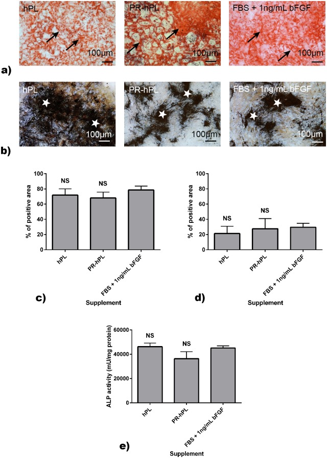
Fig 7. Osteoblast differentiation potential of BM-hMSCs after culture in an FBS+bFGF-, hPL- or PR-hPL-containing medium.
Differentiation was induced using the specific medium. The calcium deposit was stained using Alizarin Red S (a) and the extracellular matrix using Von Kossa (b). Representative photographs of experiments with hMSCs from n = 3 BM. Black arrows and white stars indicated positively stained areas. Quantification of Alizarin Red S (c) or Von Kossa (d) was expressed as a percentage of positive area. ALP activity measurement was performed using a commercially available kit (e). NS: not significant versus FBS.

