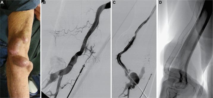
Figure 1.
Showing the angiographic technique in patient case 9.
Notes: (A) Photograph of the URL showing a native humerocephalic fistula with large venous aneurysms on the puncture site area. (B) Angiography of the URL obtained through an abbocath catheter that shows vascular access thrombosis. (C) Control fistulogram showing patency of the whole vascular access with contrast material flowing inside the stents through an intrathrombal channel: stent tunnel technique. (D) Radiography of the stents during follow-up.
Abbreviation: URL, upper right limb.

